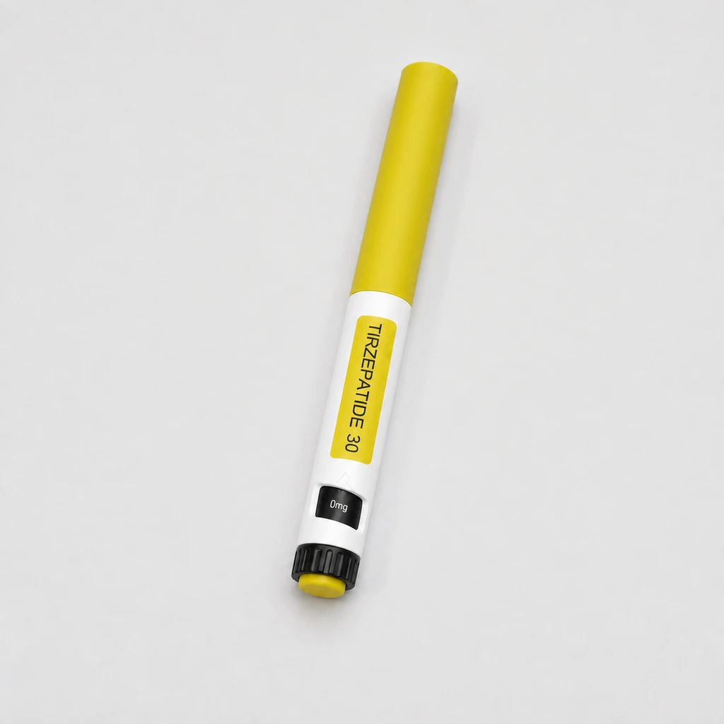
Did tirzepatide change the brain's response to every food photograph?
A headline about “rewiring food cravings” can compress an eating test, a questionnaire and a brain scan into a single claim.
A headline about “rewiring food cravings” can compress an eating test, a questionnaire and a brain scan into a single claim. A six-week tirzepatide study included different measurements of ingestive behaviour. Its imaging findings are most useful when the categories of food photos stay attached.
Which signal did the scan measure?
The randomized phase 1 study measured blood-oxygen-level-dependent activation while people viewed photographs. An fMRI BOLD response is an indirect signal associated with changes in blood oxygenation. It is not an image of a thought, a numerical appetite score or a record of what a person ate at home. The paper also assessed eating behaviour separately. Those outcomes should not be substituted for one another even when a credible biological link is possible.
What happened to the pooled category?
The investigators did not find a statistically significant change against placebo in the aggregated imaging category of highly palatable foods. They did report lower activation in specified regions for high-fat, high-sugar photos. Both statements belong together. Selecting only the positive subcategory and writing “all appetizing food images” changes the tested comparison. It also hides the importance of the multiple brain regions, categories and time points a reader may encounter in imaging work.
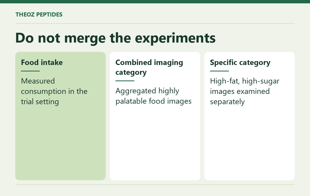
| Do not merge the experiments | What to look for |
|---|---|
| Food intake | Measured consumption in the trial setting |
| Combined imaging category | Aggregated highly palatable food images |
| Specific category | High-fat, high-sugar images examined separately |
An example away from the scanner
A photograph of a dessert, a rating of desire to eat it and the amount a person consumes at a meal can point in different directions. A Bali resident may like the appearance of a dish but decline it because a meal has just finished. A laboratory task controls parts of that context; it cannot recreate all routines, cultural preferences or food access. A brain signal alone therefore cannot identify a particular person's future food choices.
Keep three lines on the research card
Write the exact images, measured signal and comparison group on one line, the behavioral or intake results on another and the population and length of follow-up on a third. If a treatment decision is under discussion, a clinician can place those findings alongside health history rather than inferring intent or willpower from a scan. The product listing establishes a presentation; it does not turn a subset of images into a personal appetite forecast.